Exploring the Latest Diagnostic Tools: 5 Imaging Techniques for Bone Tumors

Exploring the Latest Diagnostic Tools: 5 Imaging Techniques for Bone Tumors
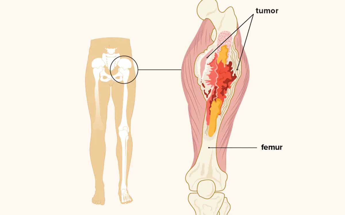
A cornerstone to efficient therapy of bone tumors, like all malignancies, is an early and precise diagnosis. Both benign and malignant bone tumors can appear with a wide range of symptoms. As a result, bone tumors are frequently discovered by chance when an individual is being examined for something completely unrelated, such as an injury.
While most bone tumors are benign, especially those with minor symptoms, diagnostic imaging is the only technique to precisely diagnose and evaluate these tumors. We'll go over the five imaging methods we use at Greater Dallas Orthopedics to make the most accurate diagnoses in this article.
X-ray
The most well-known and basic imaging tests we utilize are X-rays. They give crucial diagnostic information, and if the x-rays are of good quality, they can sometimes be the only test required to diagnose and track a benign bone tumor.
CT scan
CT scans, unlike x-rays, are a three-dimensional examination. In comparison to x-rays, these scans allow our orthopedic oncologists to see both the bone and soft tissue in far more detail, allowing them to measure bone degeneration, matrix creation, and the degree of bone involvement. CT scans are more effective than x-rays for soft tissue issues, but less effective than an MRI.
MRI
Our doctors can get incredibly detailed images of both bone and soft tissue using MRI. The use of an IV contrast, which most patients can tolerate with minimum discomfort, contributes to the MRI's effectiveness. While the image quality is excellent, the addition of a new variable in the form of the contrast media implies that the scan may need to be repeated in some circumstances to properly scan the entire area.
Bone scan
A bone scan is a nuclear medicine test that shows areas of bone turnover and is normally performed only when a diagnosis is proving problematic or to assure there are no other areas of bone involvement. In the broad sense, MRI, which we described above, has made this test considerably less important, especially in the case of benign bone cancers.
PET scan
The PET scan is a relatively new imaging technology that allows doctors to view metabolic activity within a tumor. It is most commonly used for cancer imaging, however it is occasionally used for individuals with benign bone illnesses.


